SERVICES OFFERED
ENDOSCOPY
- Diagnostic & Therapeutic Upper GI Endoscopy
- Diagnostic & Therapeutic Colonoscopy
- Capsule Endoscopy
- ERCP & Cholangio-pancreatoscopy
- Endoscopic Ultrasound [EUS]
GI MOTILITY LAB
- 24 Hrs Esophageal pH-Impedance study
- Esophageal & Anorectal Manometry
BREATH TESTS
- Hydrogen Breath Test for small intestinal bacterial overgrowth
- Urea Breath Test for H. pylori infection
SURGERY
- Esophageal & Gastric Surgery
- Hepatectomy & Liver Surgery
- Pancreato – Billiary Surgery
- Colorectal Surgery
- Hernia Surgery – Laparoscopic & Open
- Minimally Invasive & Laparoscopia Surgery
- Gl Oncology Surgery
- Surgery for Piles, Fistula & Fissure
RADIOLOGY
- Digital X-Ray
- Ultrasound with Colour Doppler
- Liver Elastography [Fibroscan] for liver fibrosis and steatosis.
PATHOLOGY
CRITICAL CARE TAIL
- State of Art Intensive Care Unit & Expert Team for management of GI and hepato-pancreato-biliary diseases
ADVANCED SERVICES OFFERED
ERCP & CHOLANGIO-PANCREATOSCOPY
- Complex Pancreato-biliary Procedures
[Multiple biliary stent placement for hilar strictures;
mechanical lithotripsy for CBO stones] - Laser Lithotripsy Treatment for CBD / pancreatic stones
- CBD Stricture evaluation
- Ampullectomy
THIRD SPACE ENDOSCOPY
- Endoscopic Mucosal Resection [EMR] & Endoscopic Submucosal Dissection [ESD] for early gastrointestinal cancers
- Peroral Endoscopic Myotomy [POEM] for Zenker’s Diverticulum,
Achalasia Cardia, Gastroparesis, Hirschsprung’s disease - Submucuosal Tunneling & Endoscopic Resection [STER]
& Endoscopic Full-Thickness Resecton (EFTR] for subepithlial tumors
ANTIREFLUX TREATMENT [FOR GERD]
- Antireflux Mucosal Ablation [ARMA]
- Endoscopic Funcoplication
ENDOBARIATRICS [FOR WEIGHT LOSS]
- Gastric Balloon placement
- Endoscopic Sleeve Gastroplasty
ENDOSCOPIC ULTRASOUND [EUS]
- EUS & EUS – guided FNAC / Biopsy for diagnosis
- EUS – guided Pancreatic Cysto-gastrostomy / Choledocho-jejunostomy / Hepatico-gastrostomy
- EUS – guided Gastric Variceal Glue & Coil treatment
What Is a CT Scan of the Abdomen?

Why Is a CT Scan Abdomen Done?
- Detailed evaluation of the liver for tumours, abscesses, cysts or cirrhosis
- Assessment of the pancreas for pancreatitis, pancreatic tumours or cysts
- Evaluating gallbladder and bile duct conditions including stones and strictures
- Investigating abdominal pain of uncertain or complex origin
- Detecting abdominal masses or swellings requiring further characterisation
- Staging of gastrointestinal cancers to assess extent and spread
- Evaluating complications of inflammatory bowel disease
- Detecting abdominal abscesses or fluid collections
- Assessing intestinal obstruction or perforation
- Guiding percutaneous drainage or biopsy procedures
- Post operative evaluation following abdominal surgery
- Monitoring known abdominal conditions and assessing treatment response
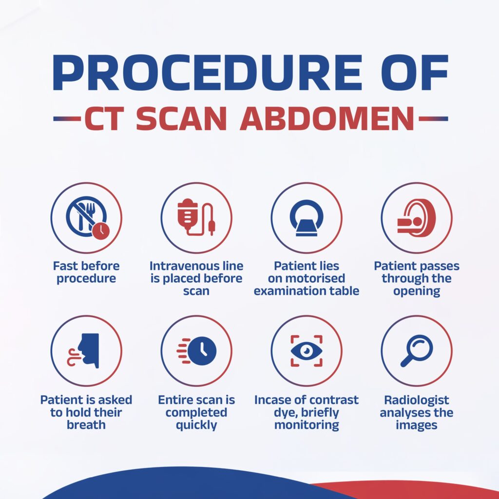
How Is a CT Scan Abdomen Performed?
- The patient may be advised to fast for a specified period before the scan depending on the clinical indication
- If contrast dye is required, an intravenous line is placed before the scan
- The patient lies on a motorised examination table that moves through the CT scanner
- The CT scanner is a large ring shaped machine and the patient passes through the opening during the scan
- The patient is asked to hold their breath briefly at certain points during the scan to obtain clear images without movement blur
- The entire scan is completed quickly and is entirely painless
- If contrast dye was administered, the patient is monitored briefly after the scan
- The radiologist analyses the images and prepares a detailed diagnostic report
What Is a Contrast CT Scan Abdomen?
What to Expect Before and After a CT Scan Abdomen
Before the procedure:
- Fasting for a specified period as instructed if contrast dye is to be administered
- Informing the radiologist or referring doctor of any known allergies, kidney conditions or current medications
- Removing metal objects including jewellery before entering the scanning room
- Wearing comfortable and loose clothing or a hospital gown as required
After the procedure:
- Normal activities can generally be resumed immediately after the scan
- If contrast dye was administered, drinking plenty of water is recommended to help the body clear the dye
- A brief observation period may be required after contrast administration in selected patients
- The radiologist will prepare a detailed report of the findings
- Results are shared with the referring gastroenterologist who will discuss findings and next steps with the patient
Why Choose LGI Hospital for CT Scan Abdomen in Nagpur?
- Specialist Radiologist with Gastroenterology Imaging Expertise: LGI Hospital in Nagpur has a dedicated radiologist with specialised expertise in abdominal and gastrointestinal imaging, ensuring accurate and comprehensive CT scan evaluation tailored to the specific needs of gastroenterology and hepatology patients.
- Advanced CT Imaging Infrastructure: LGI Hospital in Dhantoli, Nagpur is equipped with advanced CT imaging technology providing high resolution cross sectional images of the abdomen, enabling precise detection and characterisation of abdominal abnormalities.
- Seamless Integration of Imaging and Clinical Care: As a dedicated single speciality gastroenterology hospital, LGI Hospital provides seamless integration between CT imaging and clinical gastroenterology and surgical care, ensuring that scan findings are directly interpreted in the context of each patient's clinical condition.
- Accurate and Timely Reporting: LGI Hospital is committed to providing detailed, accurate and timely CT scan reports, ensuring that patients and their referring gastroenterologists receive the information needed to make prompt and well informed clinical decisions.
FAQ
Q1. Is a CT scan abdomen painful?
No, a CT scan of the abdomen is a completely painless procedure. The patient lies still on the examination table and passes through the CT scanner while images are taken. Some patients may feel a brief warm sensation if contrast dye is administered intravenously, however this is temporary and harmless. The procedure involves no physical discomfort.
Q2. How long does a CT scan abdomen take?
A standard abdominal CT scan typically takes between ten and thirty minutes including preparation time. The actual scanning process itself takes only a few minutes. Contrast CT scans may take slightly longer due to the preparation and administration of contrast dye.
Q3. What happens during a CT scan abdomen?
During a CT scan abdomen, the patient lies on a motorised table that moves through the ring shaped CT scanner. The scanner takes a series of X-ray images from multiple angles. The patient may be asked to hold their breath briefly during the scan. The radiologist analyses the cross sectional images and prepares a detailed diagnostic report. The procedure is quick and completely painless.
Q4. Why is a CT scan abdomen done?
A CT scan abdomen is performed to evaluate abdominal organs in detail including the liver, pancreas, gallbladder, spleen and intestines. It is used to diagnose conditions such as liver tumours, pancreatitis, abdominal masses, intestinal obstruction and cancer staging. It provides significantly more anatomical detail than ultrasound for complex abdominal conditions.
Q5. Which hospital is best for CT scan abdomen in Nagpur?
LGI Hospital in Dhantoli, Nagpur is a dedicated single speciality gastroenterology hospital with a specialist radiologist who performs advanced abdominal CT scanning with detailed reporting. The hospital provides seamless integration of CT imaging with clinical gastroenterology and surgical care under one roof for comprehensive abdominal evaluation.